基于CT的纵隔分区和纵隔肿瘤的鉴别诊断
CT-based mediastinal compartment classifications and differential diagnosis of mediastinal tumors.
纵隔分区有助于缩小纵隔肿瘤的鉴别诊断范围,评估肿瘤生长,并规划活检和手术。传统的几种纵隔分区法基于解剖标志和侧位胸片。最近,日本胸腺研究协会(JART)和国际胸腺恶性肿瘤兴趣小组(ITMIG)提出了基于横轴位CT图像的新的纵隔分区系统。这种基于CT的分区系统可用于更一致和精确诊断纵隔肿瘤。在本文中,我们回顾了基于CT的纵隔分区系统,并与纵隔肿瘤的鉴别诊断进行相关。
Division of the mediastinum into compartments is used to help narrow down the differential diagnosis of mediastinal tumors, assess tumor growth, and plan biopsies and surgical procedures. There are several traditional mediastinal compartment classification systems based upon anatomical landmarks and lateral chest radiograph. Recently, the Japanese Association of Research of the Thymus (JART) and the International Thymic Malignancy Interest Group (ITMIG) proposed new mediastinal compartment classification systems based on transverse CT images. These CT-based classification systems are useful for more consistent and exact diagnosis of mediastinal tumors. In this article, we review these CT-based mediastinal compartment classifications in relation to the differential diagnosis of mediastinal tumors.
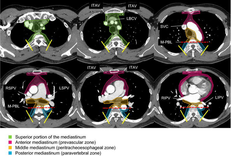
图1:轴位增强CT图像代表基于JART CT的纵隔室分类。 JART:日本胸腺研究协会; LBCV,左侧头臂静脉; ITAV,胸内动脉和静脉; SVC,上腔静脉; RSPV,右上肺静脉; LSPV,左上肺静脉; RIPV,右下肺静脉; LIPV,左下肺静脉; M-PBL,中后边界线(距胸椎椎体前缘1cm处的垂直连接)
Fig. 1 Axial contrast-enhanced CT images representing the JART CT-based mediastinal compartment classification [7]. JART: The Japanese Association for Research on the Thymus; LBCV, left brachiocephalic vein; ITAV, internal thoracic arteries and veins; SVC, superior vena cava; RSPV, right superior pulmonary vein; LSPV, left superior pulmonary vein; RIPV, right inferior pulmonary vein; LIPV, left inferior pulmonary vein; M-PBL, middle-posterior boundary line (a vertical line connecting a point on each thoracic vertebral body at 1 cm behind its anterior margin)

图2:轴位增强CT图像代表基于ITMIG CT的纵隔室分类。 ITMIG:国际胸腺恶性肿瘤兴趣小组; LBCV,左侧头臂静脉; ITAV,胸内动脉和静脉; SVC,上腔静脉; RSPV,右上肺静脉; LSPV,左上肺静脉; RIPV,右下肺静脉; LIPV,左下肺静脉; V-PBL,内脏 - 椎旁间隙边界线(距胸椎椎体前缘1cm处的垂直连接)
Fig. 2 Axial contrast-enhanced CT images representing the ITMIG CT-based mediastinal compartment classification [8]. ITMIG: The International Thymic Malignancy Interest Group; LBCV, left brachiocephalic vein; ITAV, internal thoracic arteries and veins; SVC, superior vena cava; RSPV, right superior pulmonary vein; LSPV, left superior pulmonary vein; RIPV, right inferior pulmonary vein; LIPV, left inferior pulmonary vein; V-PBL, visceral-paravertebral compartment boundary line (a vertical line connecting a point on each thoracic vertebral body at 1 cm behind its anterior margin)
Nakazono T, Yamaguchi K, Egashira R, Takase Y, Nojiri J, Mizuguchi M, Irie H. CT-based mediastinal compartment classifications and differential diagnosis of mediastinal tumors. Jpn J Radiol. 2019 Feb;37(2):117-134. doi: 10.1007/s11604-018-0777-5. Epub 2018 Sep 20. Review. PubMed PMID: 30238278.


 |手机版|小黑屋|爱科学
( 粤ICP备19015697号 )
|手机版|小黑屋|爱科学
( 粤ICP备19015697号 )


