热度 1 |||
概述:鉴别正常胸腺、胸腺增生和胸腺瘤的能力有助于重症肌无力(MG)患者的临床治疗和决策。我们试图确定常规显像在预测胸腺病理方面的准确性。
方法:我们回顾性分析牛津肌无力中心登记的接受胸腺切除术的MG患者的病历。每个病人接受1个放射学诊断和1个组织学诊断。
结果:共纳入106例患者。影像学和组织学诊断符合率为68.9%(73/73)。每种影像学诊断的敏感性和特异性分别为:胸腺瘤90%和95.5%,增生17.6%和98.6%,正常96.9%和60.8%。
讨论:常规胸部ct和MRI能有效鉴别胸腺瘤。然而,它们不是区分MG患者胸腺增生和正常胸腺的可靠工具。肌肉神经,2018。
关键词:计算机断层扫描;磁共振成像;重症肌无力;胸腺切除术;胸腺瘤;胸腺增生。
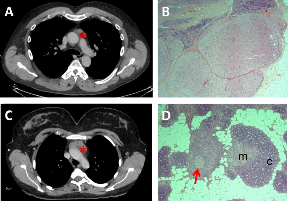
图1.2例CT扫描及胸腺组织学检查。2例患者胸部CT扫描(A,C)和胸腺组织学(B,D)的典型图像。患者1:CT扫描(A)显示前纵隔软组织(星号),可能起源于胸腺,可能代表胸腺增生。胸腺组织学(B)显示B2/B3 2期胸腺瘤的B2区,其中淋巴细胞和上皮细胞均匀混合,形成结节状边界清楚的肿块,伴有局灶性致密纤维化,取代正常胸腺结构。患者2:CT扫描(C)报告为前纵隔肿块(星号)与胸腺瘤一致。胸腺组织学(D)显示结构上保存的胸腺(胸腺皮质[c]和髓质[m]),其中有明显的淋巴滤泡和生发中心(箭头),这是滤泡增生的特征。CT,计算机断层扫描。
Fig1.CT scans and thymus histology of 2 illustrative cases. Representative images of chest CT scans (A,C) and representative images of thymus histology (B,D) for 2 distinct patients. Patient 1: CT scan (A) reported as showing anterior mediastinal soft tissue (asterisk) of likely thymic origin and likely to represent thymic hyperplasia. Thymus histology (B) illustrates B2 areas of a B2/B3, STAGE 2 thymoma in which there is an equal mix of lymphocytes and epithelial cells that form nodular well circumscribed masses with focal dense fibrosis, which replace the normal thymus architecture. Patient 2: CT scan (C) reported as having an anterior mediastinal mass (asterisk) consistent with a thymoma. Thymus histology (D) illustrates architecturally preserved thymus (thymic cortex [c] and medulla [m]) in which there are conspicuous lymphoid follicles with germinal centers (arrow), which are features of follicular hyperplasia. CT, computed tomography.
Introduction: The ability to distinguish between normal thymus, thymic hyperplasia, and thymoma should aid clinical management and decision making in patients with myasthenia gravis (MG). We sought to determine the accuracy of routine imaging in predicting thymic pathology.
Methods: We retrospectively analyzed records of patients with MG from the Oxford Myasthenia Centre registry who had undergone thymectomy. Each patient received 1 radiological diagnosis and 1 histological diagnosis.
Results: We included 106 patients. Radiological and histological diagnoses agreed in 73 (68.9%) patients. Sensitivity and specificity, respectively, were calculated for each radiological diagnosis as follows: thymoma 90% and 95.5%, hyperplasia 17.6% and 98.6%, and normal 96.9% and 60.8%.
Discussion: Routine chest computed tomography and MRI can effectively identify thymoma. However, they are not reliable tools to differentiate between thymic hyperplasia and normal thymus in patients with MG. Muscle Nerve, 2018.
Keywords: computed tomography; magnetic resonance imaging; myasthenia gravis; thymectomy; thymoma; thymus hyperplasia.


 |手机版|小黑屋|爱科学
( 粤ICP备19015697号 )
|手机版|小黑屋|爱科学
( 粤ICP备19015697号 )
GMT+8, 2025-10-4 02:11 , Processed in 0.160387 second(s), 29 queries .
Powered by Discuz! X3.5
© 2001-2013 Comsenz Inc.


