Behçet病前纵隔脂肪的CT定性定量分析
Anterior mediastinal fat in Behçet's disease: qualitative and quantitative CT analysis
富脂肪的前纵隔可作为监测系统性血管炎血管微小变化的敏感窗口。为了评估这一假设,我们对Behçet病患者和对照组的纵隔前脂肪进行了分析。本研究包括43例在过去11年内确诊为Behçet病的患者,他们接受了CT扫描;选择55例患者作为对照组。根据CT表现对纵隔脂肪进行分类。比较血清炎症标志物,根据形态类型评价疾病活性,并测定前纵隔平均HU。Behçet病组的平均CT值明显高于对照组(-48.5±33.5 vs.-67.7±18.7),P<0.05。纵隔脂肪类型分为:单纯脂肪组织(2例与31%(Behçet病与对照组比较),弥漫性软组织浸润(16 vs.29%),管状结构(21 vs.4%),混合浸润与管状结构(42 vs.15%),以及明显胸腺组织(19 vs.22%)。C反应蛋白水平高的组纵隔平均CT值明显高于正常组。Behçet病组纵隔前脂肪的平均CT值明显高于正常组。尽管需要病理证实,但其原因可能是由Behçet病引起的炎症性新生血管或胸腺增生。
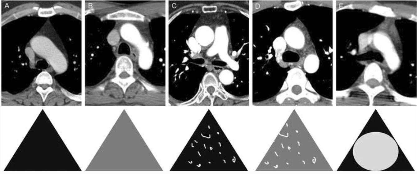
The fat-rich anterior mediastinum could be a sensitive window for monitoring minute changes in vascularity induced by systemic vasculitis. To evaluate this hypothesis, an analysis of anterior mediastinal fat in patients with Behçet's disease and a control group was conducted. This study included 43 patients diagnosed with Behçet's disease within the last 11 years who underwent CT scan; 55 patients were selected as a control population. Mediastinal fat was classified according to CT morphology. Comparison of serum inflammatory markers was performed for evaluation of disease activity according to morphologic types, and average Hounsfield unit of the anterior mediastinum was measured. Significantly higher mean CT attenuation was observed in the Behçet's disease group, compared with the control group (-48.5 ± 33.5 vs. -67.7 ± 18.7, respectively, P < 0.05). Mediastinal fat types were classified as follows: pure fatty tissue (2 vs. 31 % [Behçet's disease vs. control group]), diffuse soft tissue infiltration (16 vs. 29 %), tubular structures (21 vs. 4 %), mixed infiltration with tubular structures (42 vs. 15 %), and evident thymic tissue (19 vs. 22 %). The value for mean mediastinal attenuation was significantly higher in the group with a high level of C-reactive protein than in the normal level group. The mean CT attenuation of anterior mediastinal fat is significantly higher in the Behçet's disease group, compared with the normal group. Although pathologic confirmation is needed, the cause is postulated to be either inflammatory neovascularization or minimal thymic hyperplasia induced by Behçet's disease.


 |手机版|小黑屋|爱科学
( 粤ICP备19015697号 )
|手机版|小黑屋|爱科学
( 粤ICP备19015697号 )


