重症肌无力患者正常和淋巴滤泡增生胸腺CT对比研究
Comparative study of computed tomography of normal and lymphoid follicular hyperplasia thymus in myasthenia gravis patients
本研究的目的是评估非胸腺瘤性重症肌无力(MG)患者的胸腺,通过CT来区分淋巴滤泡增生(LFH)胸腺与正常/退化胸腺,以帮助外科医生确定非胸腺瘤性MG患者是否需要手术。
本文回顾性分析了天津医科大学附属总医院(天津,中国)80例接受CT扫描和胸腺切除术的患者。经病理证实,CT初诊54例为LFH胸腺。胸腺测量,包括前后和横向径线、宽度(横轴位图像胸腺叶的最长轴)和厚度(垂直于胸腺叶长轴的最大径线)以及每个CT切片中胸腺区域、脂肪组织和胸壁肌肉组织的CT值,以评估在LFH组和正常/退化胸腺组之间的差异。
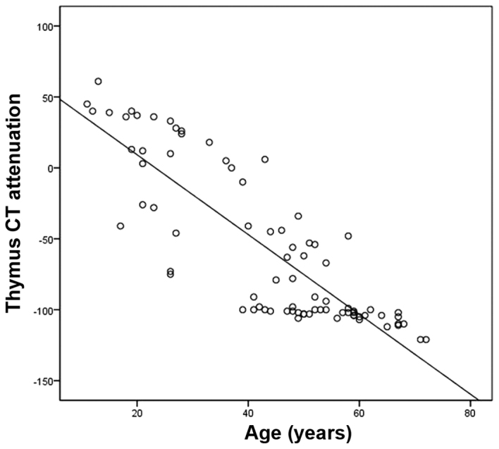
尽管患者年龄与胸腺区域CT值呈负相关(r=-0.779,P<0.05,Pearson相关检验),但LFH胸腺组在CT上表现为结节性改变,而正常/退化胸腺组未观察到此类改变。LFH胸腺组的平均发病年龄明显低于正常胸腺组(40.2±17.3岁,59.2±9.3岁)。此外,LFH组和正常/退化胸腺组之间的CT值存在显著差异[未增强CT:-41.21±54.42 vs.-108.23±8.72 HU;增强CT:-25.57±58.65 vs.-117.40±6.22 HU]。在LFH组,胸腺区域和脂肪组织之间的平均CT值存在显著差异,而在正常/退化胸腺组中没有观察到显著差异。
结论:CT可用于MG患者LFH胸腺与正常/退化胸腺的鉴别。
图1.胸腺CT值与年龄的相关性(r=−0.779;P<0.05)。
Figure 1. Correlation of the thymus CT attenuation with the age of the patients (r=−0.779; P<0.05). CT, computed tomography.
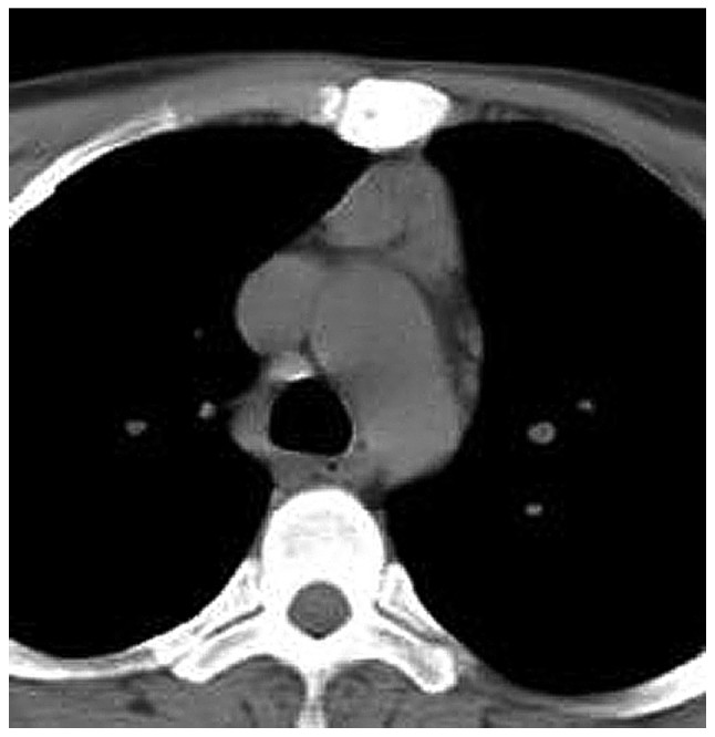
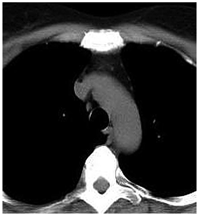
图2. 男性重症肌无力患者(年龄18岁)淋巴滤泡增生的非增强CT图像。胸腺呈三角形,很容易与邻近组织区分开。
Figure 2. Unenhanced computed tomography image of a male myasthenia gravis patient (age, 18 years) with lymphoid follicular hyperplasia. The thymus is triangular in shape and may be easily distinguished from adjacent tissue.
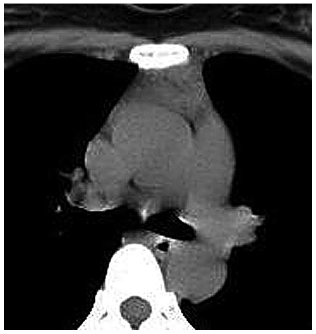
图3. 女性重症肌无力患者(年龄26岁)伴淋巴滤泡增生。胸腺双侧叶之间可见间隔。
Figure 3. Female myasthenia gravis patient (age, 26 years) with lymphoid follicular hyperplasia. The septum between the two lobes is visible.
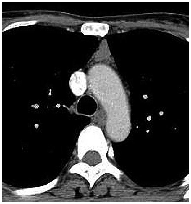
图4. 女性重症肌无力患者(年龄36岁),淋巴滤泡增生。胸腺呈四边形,胸腺区的CT值与脂肪组织和肌肉不同。
Figure 4. Female myasthenia gravis patient (age, 36 years) with lymphoid follicular hyperplasia. The thymus is quadrilateral and the computed tomography attenuation in the thymus region is different from that of the adipose tissue and musculature.
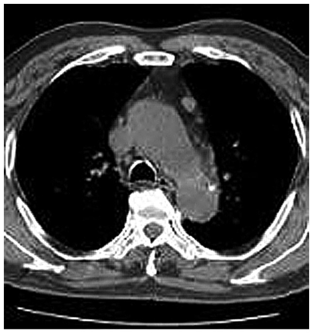
图5. 女性重症肌无力患者(年龄45岁)伴淋巴滤泡增生。胸腺呈四边形,脂肪浸润区可见结节。
Figure 5. Female myasthenia gravis patient (age, 45 years) with lymphoid follicular hyperplasia. The thymus is quadrilateral in shape and nodules are visible in the fat-infiltrated thymus region.
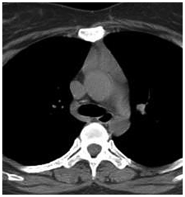
图6. 一例女性重症肌无力患者(年龄50岁)淋巴滤泡增生的增强CT图像。胸腺呈三角形,很容易与邻近组织,特别是脂肪组织区分开来。
Figure 6. Contrast-enhanced computed tomography image of a female myasthenia gravis patient (age, 50 years) with lymphoid follicular hyperplasia. The thymus is triangular in shape and can be easily distinguished from adjacent tissue, particularly adipose tissue.
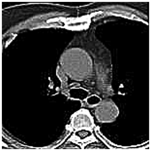
图7. 女性重症肌无力患者(年龄,54岁)伴LFH。胸腺呈三角形,可见结节,病理证实为LFH。淋巴滤泡增生。
Figure 7. Female myasthenia gravis patient (age, 54 years) with LFH. The thymus is triangular in shape and a nodule is apparent, which was later pathologically confirmed as LFH. LFH, lymphoid follicular hyperplasia.
图8.女性重症肌无力患者(年龄58岁)淋巴滤泡增生的CT增强扫描。胸腺呈三角形,胸腺区可见结节。
Figure 8. Contrast-enhanced computed tomography image of a female myasthenia gravis patient (age, 58 years) with lymphoid follicular hyperplasia. The thymus is triangular in shape and nodules are apparent in the thymus region.
图9. 一例男性重症肌无力和淋巴滤泡增生患者(年龄67岁)的CT平扫图像。胸腺呈四边形,某些结节可与脂肪组织区分开。
Figure 9. Unenhanced computed tomography image of a male patient (age, 67 years) with myasthenia gravis and lymphoid follicular hyperplasia. The thymus is quadrilateral in shape and certain nodules may be distinguished from adipose tissue.
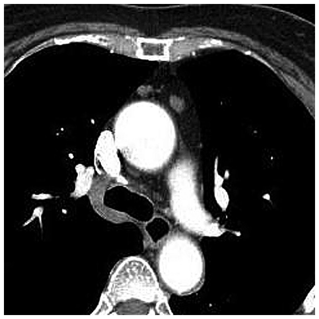
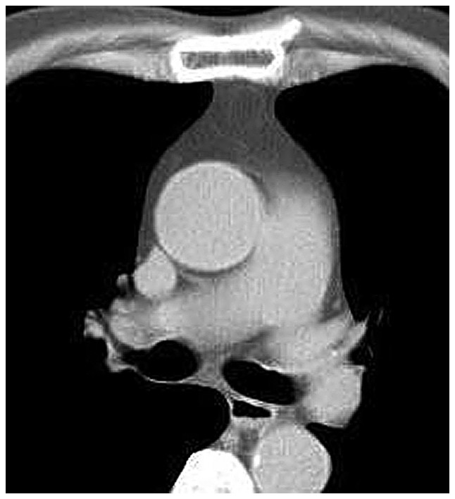
图10. 一例男性重症肌无力(43岁)正常胸腺的CT增强增强图像。胸腺呈四边形,CT值均匀。
Figure 10. Contrast-enhanced CT image of a male myasthenia gravis patient (age, 43 years) with normal thymus. The thymus is quadrilateral in shape and homogeneous regarding CT attenuation. CT, computed tomography.
图11. 男性重症肌无力患者(年龄59岁)正常胸腺(退化)的CT平扫图像。胸腺呈四边形,CT值均匀。
Figure 11. Unenhanced CT image of a male myasthenia gravis patient (age, 59 years) with normal (involuted) thymus. The thymus is quadrilateral in shape and homogeneous regarding CT attenuation. CT, computed tomography.
图12.一例胸腺正常的男性重症肌无力患者(年龄72岁)的CT平扫图像。胸腺呈四边形,CT值均匀。
Figure 12. Unenhanced CT image of a male myasthenia gravis patient (age, 72 years) with normal thymus. The thymus is quadrilateral in shape and homogeneous regarding CT attenuation. CT, computed tomography.
The aim of the present study was to evaluate the thymuses of non-thymomatous myasthenia gravis (MG) patients by computed tomography (CT) for differentiating lymphoid follicular hyperplasia (LFH) thymus from normal/involuted thymus in order to assist surgeons in determining whether a non-thymomatous MG patient requires an operation. In the present retrospective review over 10 years, 80 patients who received CT scan and thymectomy at the Affiliated General Hospital of Tianjin Medial University (Tianjin, China) were included. According to the pathological records, 54 of the cases initially detected on CT were confirmed as LFH thymus. Thymic measurements, including anteroposterior and transverse dimensions, width (the longest axis of the lobe on a transverse scan) and thickness (the largest dimension perpendicular to the long axis of the lobe) and CT attenuation of the thymus region, adipose tissue and chest wall musculature in each CT slice were included to assess differences between the LFH group and the normal/involuted thymus group. Although a negative association between patient age and the CT attenuation of the thymus region was identified (r=-0.779, P<0.05, Pearson's correlation test), the LFH thymus group featured nodular changes on CT, while no such changes were observed in the normal/involuted thymus group. The mean age of disease onset in the LFH thymus group was significantly lower than that in the normal thymus group (40.2±17.3 vs. 59.2±9.3 years). Furthermore, significant differences in CT attenuation were identified between the LFH group and the normal/involuted thymus group [-41.21±54.42 vs. -108.23±8.72 Hounsfield units (HU) on unenhanced CT; -25.57±58.65 vs.-117.40±6.22 HU on contrast-enhanced CT]. In the LFH group, the difference in mean CT attenuation between the thymus region and adipose tissue was significant, while no significant difference was observed in the normal/involuted thymus group. In conclusion, CT may be used to distinguish LFH thymus from normal/involuted thymus in MG patients.
Zhang H, Zhang P, Yu TL. Comparative study of computed tomography of normal and lymphoid follicular hyperplasia thymus in myasthenia gravis patients. Exp Ther Med. 2019 Jan;17(1):512-518. doi: 10.3892/etm.2018.6948. Epub 2018 Nov 9. PMID: 30651830; PMCID: PMC6307392.


 |手机版|小黑屋|爱科学
( 粤ICP备19015697号 )
|手机版|小黑屋|爱科学
( 粤ICP备19015697号 )


