儿童胸腺:识别正常和异位胸腺组织。The paediatric thymus: recognising normal and ectopic thymic tissue.
儿童胸腺的出现随着胸腺退化的正常过程而改变。 胸腺组织可以在前纵隔内原位,也可以在胚胎发育过程中位于异位。 儿童影像学检查中原位和异位胸腺组织形态的改变可能会导致将正常胸腺误认为是病变。 正常胸腺组织的识别可以降低不必要的进一步诊断检查和患者焦虑。 在这篇综述中,我们讨论了正常胸腺的胚胎发育和解剖学变异,并展示了儿童正常胸腺的多模态影像特征,包括正电子发射断层扫描,扩散加权成像以及同反相位磁共振成像。 我们展示了正常的假性胸腺病变,并从病理学角度讨论了区分正常胸腺的特征,包括胸腺反弹性增生。
The appearance of the paediatric thymus changes as the normal process of thymic involution occurs. Thymic tissue may be orthotopic within the anterior mediastinum or ectopically located along the course of its embryological development. The variable appearance of orthotopic and ectopic thymic tissue in children on imaging studies may lead to misinterpretation of the normal thymus as pathology. Recognition of normal thymic tissue can mitigate unnecessary further diagnostic testing and patient anxiety. In this review, we discuss the embryological development and anatomical variants of normal thymus, and demonstrate the multimodality imaging features of the normal thymus in children, including positron-emission tomography, and diffusion-weighted imaging and in- and opposed-phase imaging on magnetic resonance imaging. We demonstrate the normal thymus mimicking pathological processes and discuss features that distinguish normal thymus, including thymic rebound hyperplasia, from pathology.

图1.(a)胎龄6周时胸腺发育。 两侧的第三咽袋(粉红色附属物)形成胸腺原基,成为胸咽管。 导管尾部和内侧下降(紫色箭头),在前纵隔融合。 (b)异位胸腺的正常变异位置(绿色结构)可以位于咽喉导管下降路径的任何位置,包括下颌下和甲状腺内位置。 正常的胸腺也可以像颈状胸腺一样延伸到胸骨柄(Manubrium)上方,紧靠甲状腺。
Figure 1. (a) Thymus development at 6 weeks gestational age. The bilateral third pharyngeal pouches (pink appendages) form the thymic primordia, which become the thymopharyngeal ducts. The ducts descend caudally and medially (purple arrows) to fuse at the anterior mediastinum. (b) Normal variant locations of ectopic thymus (green structures) can be anywhere along the pathway of descent of the thymopharyngeal duct, including submandibular and intrathyroidal locations. The normal thymus can also extend above the manubrium as a cervical thymus, abutting the thyroid gland.

图2.新生儿胸片上的正常胸腺。 (a)一名2个月大的男性患儿,胸腺突出,显示“帆征”(箭头)。 还可以看到“波浪形”(箭头)。 (b)新生儿,在心脏轮廓上有原位胸腺,形成心脏肿大的外观。 出现“凹痕征 ”(箭头)表示胸腺与心脏交接的下边界。 男性新生儿可见较大的纵隔气肿,胸腺抬高并在(c)侧位片和(d)额片上产生“三角帆征”(箭头)。
Figure 2. Normal thymus on chest radiographs of neonates. (a) A 2-month-old male patient with a prominent thymus displaying the “sail sign” (arrow). The “wave sign” (arrowhead) is also seen. (b) Female neonate with the orthotopic thymus over the cardiac silhouette creating the appearance of cardiomegaly. The presence of the “notch sign” (arrow) delineates the inferior border of the thymus from the heart. Male neonates with a large pneumomediastinum, elevating the thymus and producing the “spinnaker sign” (arrows) on (c) lateral and (d) frontal radiographs.
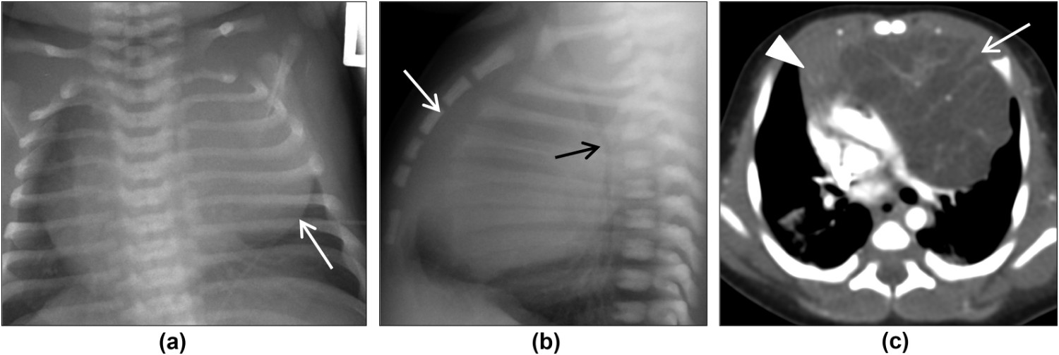
图3.刚出生时出现咕哝和呼吸窘迫的男性新生儿。 (a)正位和(b)侧位胸部X线片显示前纵隔软组织肿块(白色箭头)导致气管向后移位和受压(黑色箭头)。(c)轴位增强CT图像显示胸腺左叶(箭头)可见混杂多囊性囊性肿块,纵隔血管结构受压向后和向右移位。右前纵隔可见正常的胸腺组织(箭头)。组织学证实肿块是生殖细胞肿瘤。
Figure 3. Male neonate presenting with grunting and respiratory distress at birth. (a) Frontal and (b) lateral chest radiographs demonstrate anterior mediastinal soft-tissue mass (white arrows) causing posterior displacement and compression of the trachea (black arrow). (c) Transverse contrast-enhanced CT image demonstrates a heterogeneous multiseptate cystic mass arising from left lobe of thymus (arrow), displacing mediastinal vascular structures posteriorly and to the right. Normal thymic tissue is present in the right anterior mediastinum (arrowhead). Histology confirmed mass to be a germ cell tumour.

图5.胸部CT上的正常原位胸腺。 (a)一名6个月大女性患儿,可见明显的双凸型正常胸腺(箭头),轴位增强CT图像。 (b)一名11岁男性患儿的轴位增强CT图像显示部分凹陷型原位胸腺,外缘边界平直(箭头)。
Figure 5. Normal orthotopic thymus on chest CT. (a) Contrast-enhanced transverse CT image of a 6-month-old female patient with a prominent biconvex normal thymus (arrows). (b) Transverse image from contrast-enhanced CT in an 11-year-old male patient shows partially involuted orthotopic thymus with straight borders (arrows).
图6.15岁女性患者,疲劳和虚弱。 (a)正位和(b)侧位胸部X线片显示左前纵隔肿块(箭头)。(c)横断面增强CT图像显示,由于胸腺左叶(箭头)向后移位左肺动脉并导致左肺上叶不张形成不均质软组织肿块。 胸腺的右叶(箭头)的大小和轮廓随年龄增长而变化。 组织学证实肿块为胸腺瘤。
Figure 6. A 15-year old female patient with fatigue and weakness. (a) Frontal and (b) lateral chest radiographs demonstrate a left anterior mediastinal mass (arrows). (c) Transverse contrast-enhanced CT image demonstrates a heterogeneous soft-tissue mass arising from left lobe of thymus (arrow) displacing the left pulmonary artery posteriorly and causing atelectasis in the left upper lobe. The right lobe of thymus (arrowhead) has normal size and contour for age. Histology confirmed mass to be thymoma.
图7. 4.5岁男性患者的胸部MRI排除了胸腺瘤的存在。 MRI上原位胸腺的冠状位图像,向颈部延伸代表颈位胸腺。(a)原位胸腺的T1WI显示相对肌肉呈轻度高信号,而相对脂肪则呈低信号。 (b)脂肪饱和T2WI显示原位胸腺相对于肝脏和肌肉呈轻度高信号,并且在脂肪饱和序列上并未饱和,提示为儿童胸腺组织。 (c)胸部的轴位DWI(b = 800)图像以及(d)相应的ADC图表明正常原位胸腺的扩散不受限。
Figure 7. Chest MRI of 4.5-year-old male patient to exclude the presence of thymoma. Coronal images of the orthotopic thymus on MRI with cervical extension representing a cervical thymus. (a) T1WI of the orthotopic thymus shows that it is mildly hyperintense to muscle and hypointense to fat. (b) T2WI with fat saturation shows the orthotopic thymus is mildly hyperintense to the liver and the muscle, and does not saturate on fat saturation sequence, as expected of the paediatric thymus. (c) Transverse DWI (b = 800) image of the chest with the (d) corresponding ADC map indicates unrestricted diffusion of the normal orthotopic thymus.

图8.12岁女性患儿,慢性咳嗽。 (a)正位胸片显示纵隔增宽(箭头),高度怀疑淋巴瘤。 增宽的纵隔(箭头)行反相(b)和同相(c)T1WI检查,在反相位图像中可见纵隔前部组织的信号强度出现轻度降低。 活检证实胸腺增生。
Figure 8. A 12-year-old female patient who was being investigated for chronic cough. (a) Frontal chest radiograph shows a widened mediastinum (arrowheads), raising concerns for lymphoma. Opposed (b) and in-phase (c) T1WI performed for work-up of the widened mediastinum (arrow) revealed mild signal intensity loss of the anterior mediastinal tissue on opposed-phase imaging. Biopsy confirmed thymic hyperplasia.
Wee T, Lee AF, Nadel H, Bray H. The paediatric thymus: recognising normal and ectopic thymic tissue. Clin Radiol. 2021 Mar 21:S0009-9260(21)00124-0. doi: 10.1016/j.crad.2021.02.017. Epub ahead of print. PMID: 33762135.


 |手机版|小黑屋|爱科学
( 粤ICP备19015697号 )
|手机版|小黑屋|爱科学
( 粤ICP备19015697号 )


