|
|
横膈,也叫膈肌,横膈膜,diaphragm,是胸部(thorax)和腹部(abdomen)的分界,是胸腔的底和腹腔的顶。横膈是向上膨隆的组织,周围是肌肉,就是膈肌,中心是腱膜,就是中心腱(central tendon)。膈肌是横纹肌(striated muscle),起自胸廓下口周缘,前方与胸骨连接,两侧与肋骨连接,后方与脊椎贴近,终止于中心腱。横膈(diaphragm)与纵隔(mediastinum)均是中文翻译,表达了这种分隔组织的纵横位置,其实二者的结构与功能完全不同。Diaphragm,来自拉丁语 diaphragma,词源希腊语 diaphragma,"partition, barrier, muscle which divides the thorax from the abdomen,"。Diaphrassein "to barricade,"分隔, 前缀 dia- "across" 横过,phrassein "to fence or hedge in."包围。
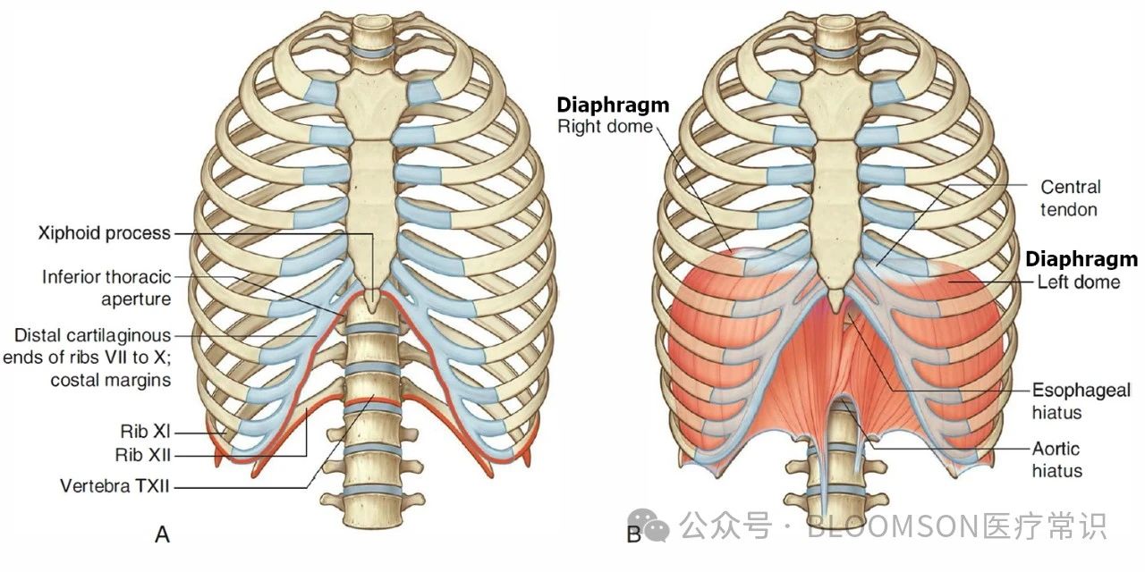
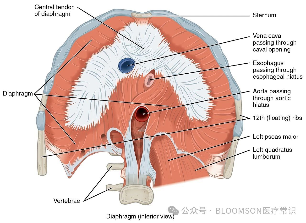
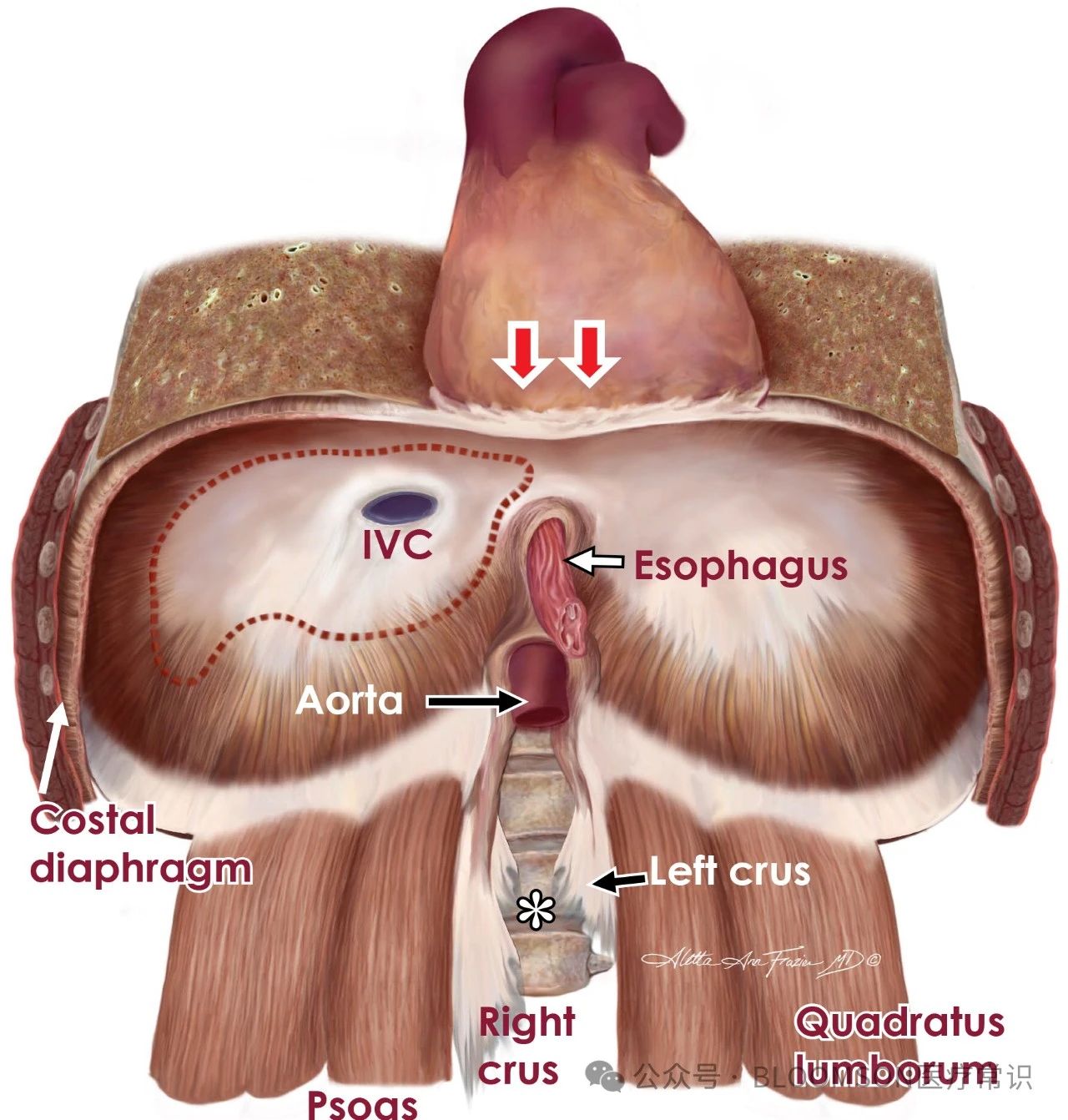
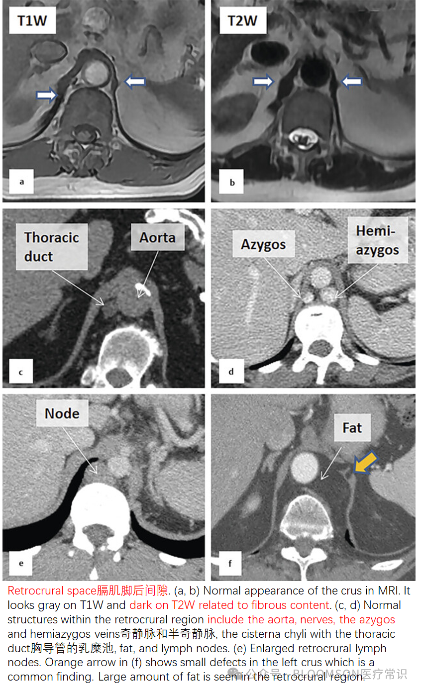
横隔上有三个裂孔,分别是主动脉裂孔(主动脉与胸导管通过),食管裂孔(食管与迷走神经通过),腔静脉裂孔(下腔静脉通过)。也就是说,食管,降主动脉,下腔静脉分别有不同的裂孔穿越横膈,其中下腔静脉(inferior vena cava)穿越了中心腱。
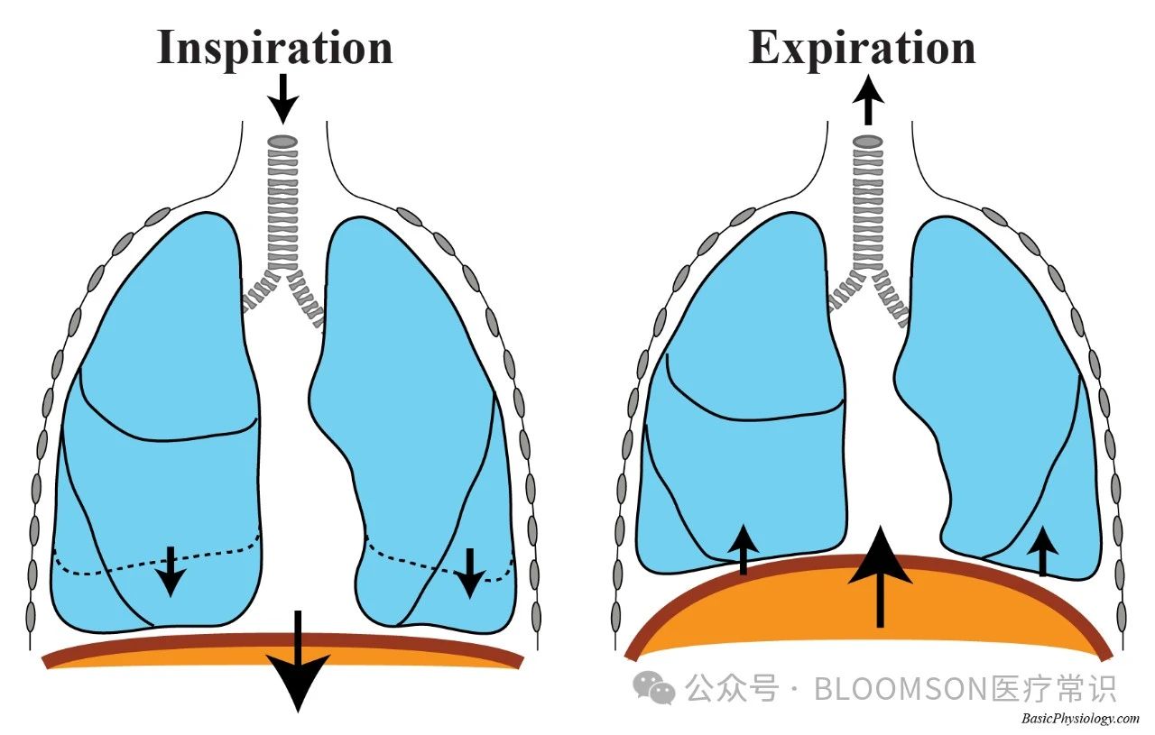
膈肌参与呼吸运动,膈肌舒张时位置上抬,右侧靠近肝脏,左侧靠近胃、脾,形成两个穹窿,当膈肌收缩时位置下落,收缩越强,膈位置越低,胸腔上下径越长,胸腔容积越大。
一、胸膜与横膈壁胸膜的组成部分包括横膈部分(Diaphragmatic part)。
The parietal pleura is divided into three subdivisions:Mediastinal part - covers the mediastinum and its structures; 纵隔胸膜Costal part - covers the inner surface of the thoracic cage including the ribs;肋胸膜Diaphragmatic part - covers the diaphragm.横膈胸膜

The pleura divides into:visceral pleura which covers the surface of the lung and dips into the fissures between its lobesparietal pleura which lines the inner of the chest wall and named according to the site it lines:cervical pleura 颈胸膜costal pleura 肋胸膜diaphragmatic pleura 横膈胸膜mediastinal pleura 纵隔胸膜
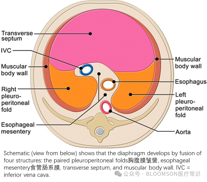
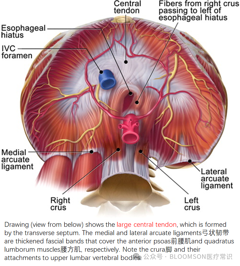
二、腹膜与横膈
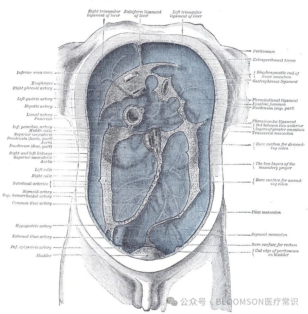
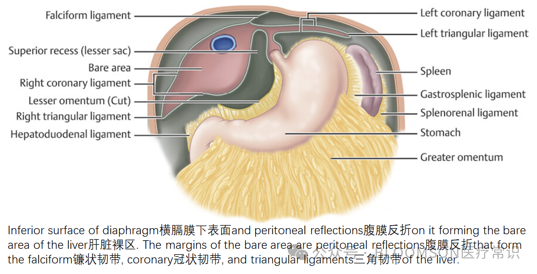
肝韧带是腹膜的双层折叠,将肝脏连接到周围器官或腹壁上。大多数与肝脏相关韧带是胚胎血管的残余,随着胎儿的发育而退化。Anterior coronary ligament of liver, 肝前冠状韧带,或者Superior part of coronary ligament of liver,肝上部冠状韧带位于横隔下方。Liver ligaments are double-layered folds of peritoneum that attach the liver to surrounding organs, or to the abdominal wall. The majority of ligaments associated with the liver are remnants of embryological blood vessels that regressed as the fetus developed.
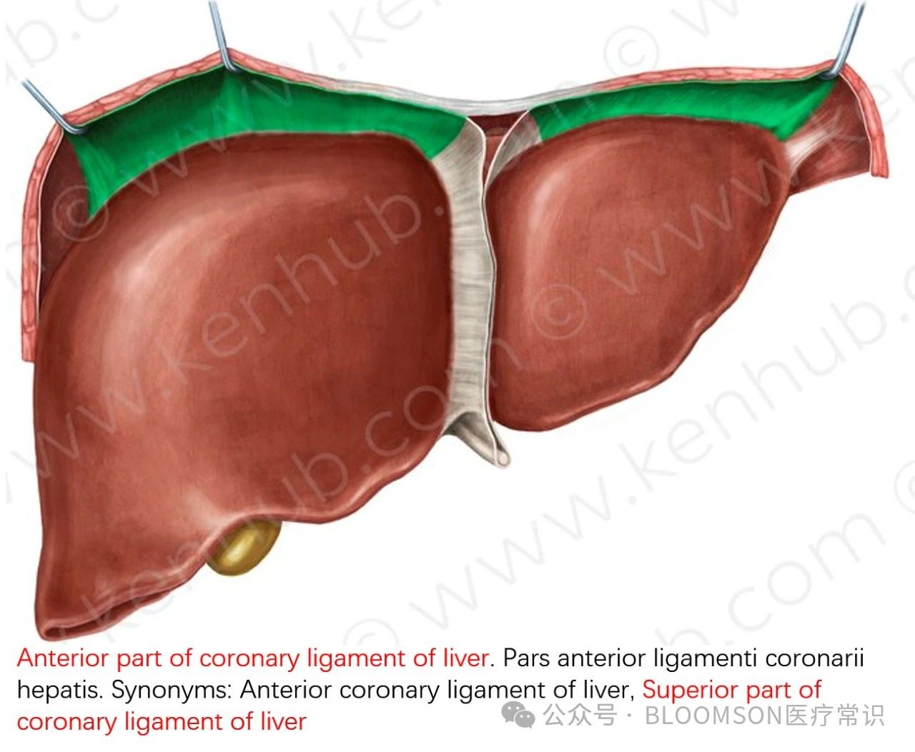
冠状韧带是肝脏的腹膜韧带复合体,包围着肝脏的裸露区域。冠状韧带是腹膜的反折,从横膈膜下的腹膜到肝脏右叶的上面和后面。肝脏的冠状韧带由上下两层组成,容纳了下腔静脉和右肾上腺的上方,一个大致呈三角形的区域。The coronary ligament is a peritoneal ligament complex of the liver which encloses the bare area of the liver. The coronary ligament is formed by the reflection of the peritoneum from the undersurface of the diaphragm onto the superior and posterior surfaces to the right lobe of the liver. It is made up of a superior and inferior layer. The superior and inferior reflections enclose the bare area of the liver, a roughly triangular area, that houses the IVC and the superior aspect of the right adrenal gland.
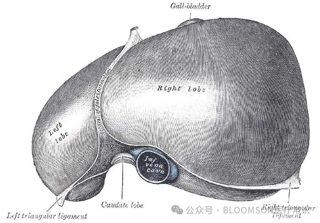
三、横膈的CT影像
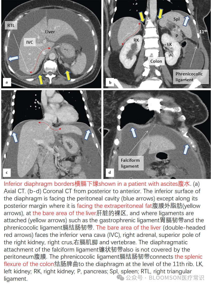
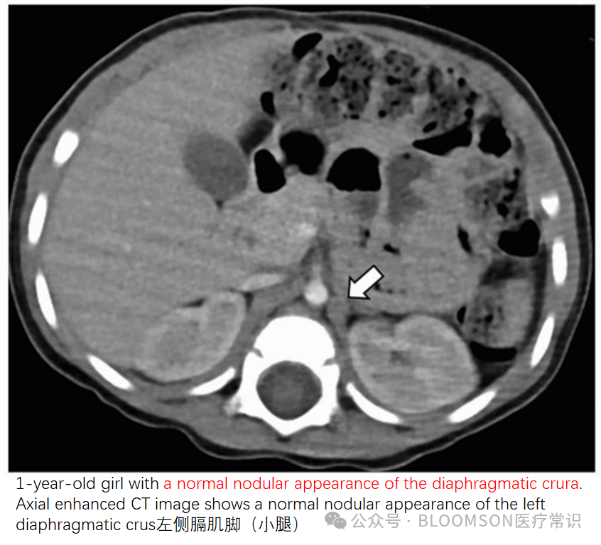
横断面CT影像,有时候难以分辨淋巴结或者病灶与横膈的关系,是膈上(胸腔),还是膈下(腹腔)。冠状面CT影像可能有助于辨别。
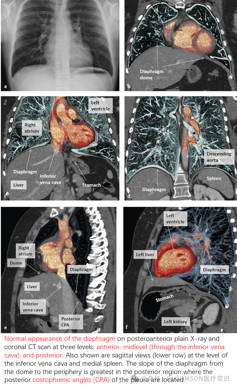
四、心膈角,cardiophrenic angle心膈角,心膈间隙,cardiophrenic space,是一个泛指,可能源于X线片的概念,是一个充满脂肪的空间,在心脏个膈肌之间。起源于膈上或膈下的病变表现为心膈角的病变。X线的概念也有肋膈角(costophrenic angles),变钝提示胸腔积液。
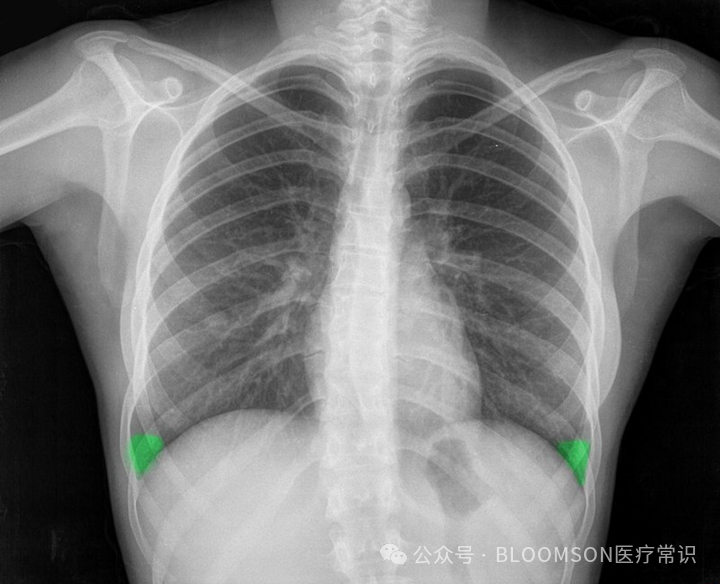
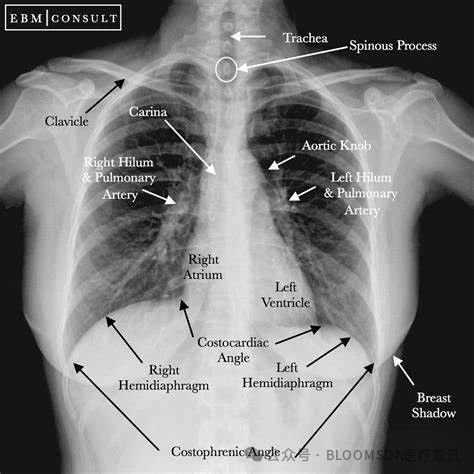
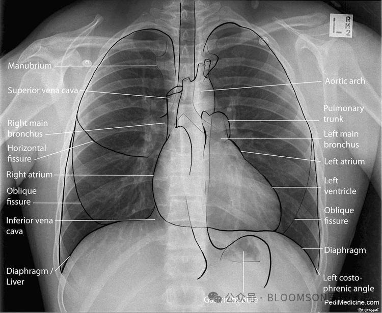
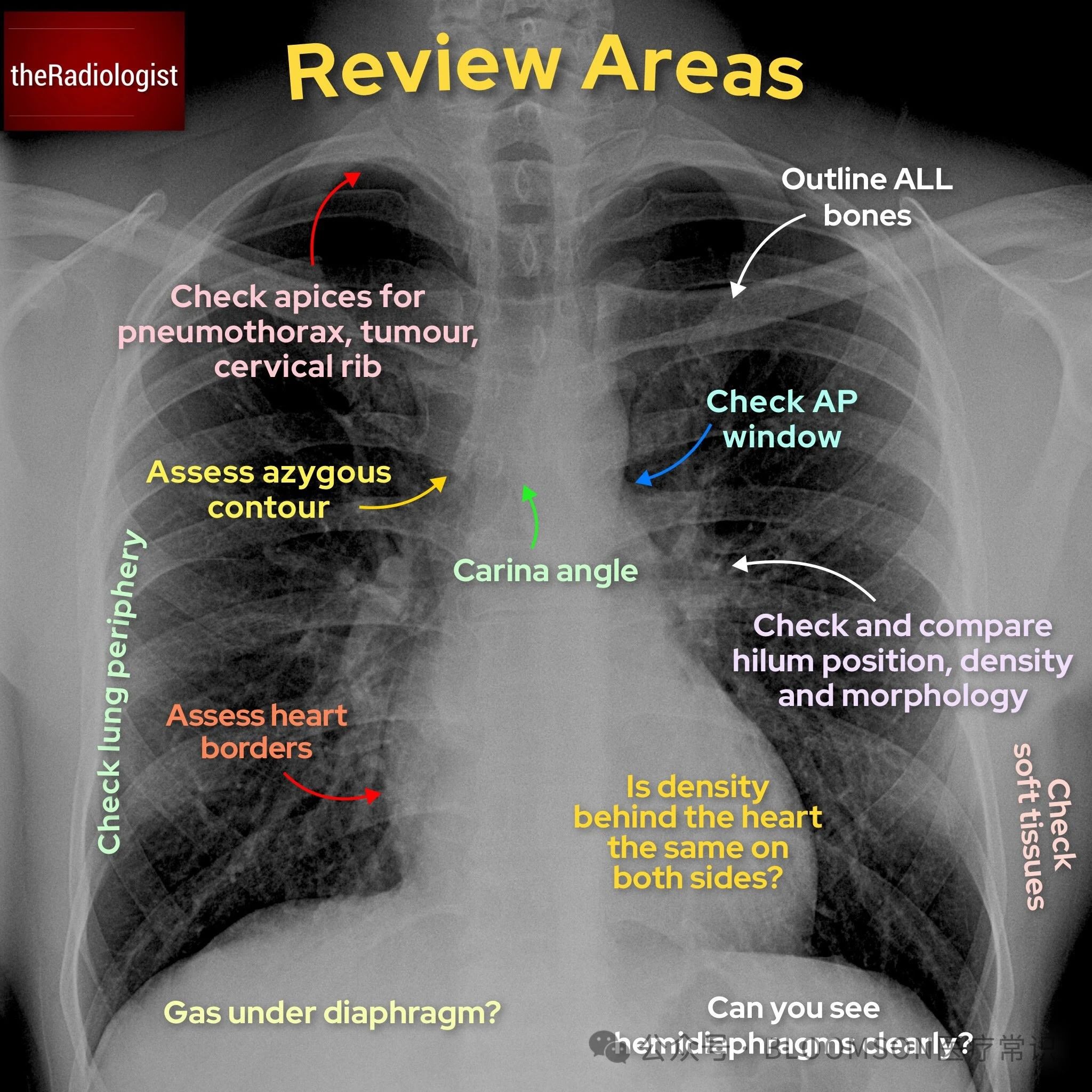
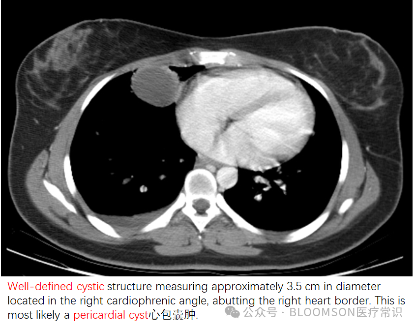
The cardiophrenic space is usually filled with fat. However, lesions originating above or lower to the diaphragm can present as cardiophrenic angle lesions.心膈角的病变包括:pericardial fat pad 心包脂肪垫pericardial cyst 心包囊肿pericardial fat necrosis 心包脂肪坏死Morgagni's hernia Morgagni's疝 lymphadenopathy: metastasis, lymphoma, reactional 淋巴结病: 转移,淋巴瘤,反应性pericardial lipomatosis 心包脂肪增多症neurogenic tumor 神经源性肿瘤thymoma 胸腺瘤right middle lobe collapse 右肺中叶塌陷right middle lobe consolidation 右肺中叶实变 impending cardiac volvulus: an abnormal bulging can be seen at the cardiophrenic angle, preceding cardiac volvulus 心肌扭转前可见心肌角异常隆起fibrous tumor of the pleura 胸膜纤维性肿瘤hydatid cyst 包虫囊varices and dilated pericardiacophrenic veins 静脉曲张和扩张的心包静脉
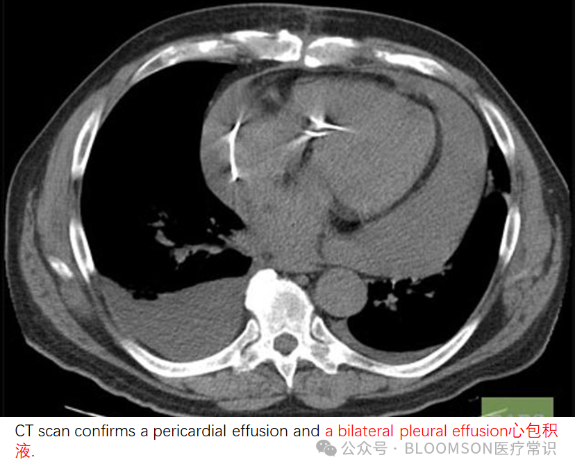
Fat collections in the right cardiophrenic angle include an enlarged epicardial fat pad, lipoma, thymolipoma, or Morgagni hernia with mesenteric fat and are the most common causes of a mass in this area.
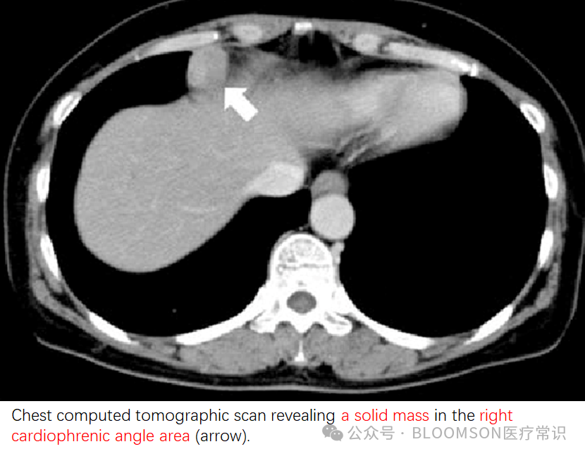
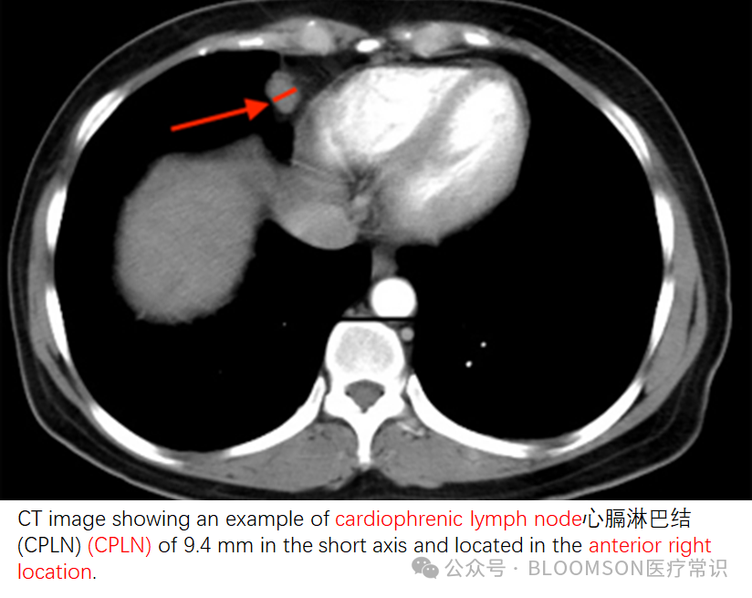
心膈角淋巴结,Cardiophrenic lymph nodes,似乎也是含糊的概念,有定义是位于心包前方(pericardium)的距离膈肌2厘米以内的淋巴结,也叫心包前淋巴结(anterior prepericardiac nodes),负责通过内乳淋巴链(internal mammary chain)引流膈肌、胸膜、腹壁前壁和肝脏。
Cardiophrenic lymph nodes were defined as those lymph nodes located anterior to the pericardium and within 2 cm of the diaphragm. These have alternatively been described as anterior prepericardiac nodes and are responsible for draining the diaphragm, pleura, anterior abdominal wall, and liver through the internal mammary chain.
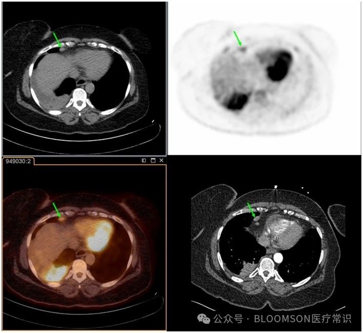

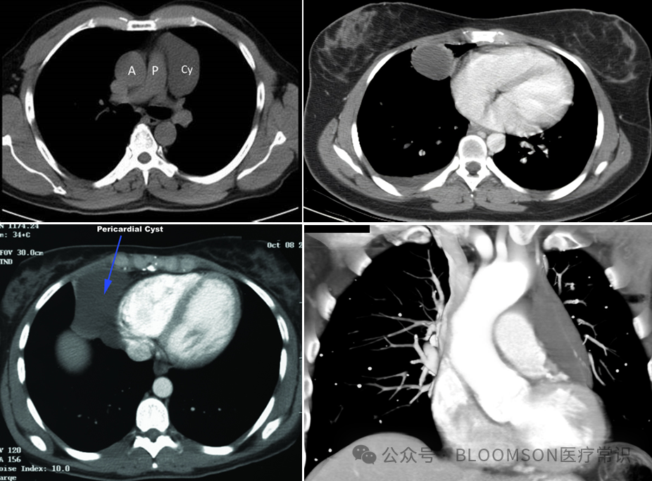
心包囊肿(pericardial cyst)最常见的部位是右侧心肌角,与脂肪垫相比,心包囊肿为组织不透明。但左侧心肌角可见任何一种病变。
来源:BLOOMSON医疗常识
|
|